

A Clinical Reference for Avian Practitioners | Harrison’s Bird Foods
Bianca Murphy, DVM, DABVP (Avian Practice)
Introduction
Atherosclerosis is one of the most common and most underdiagnosed conditions in companion psittacines. It progresses silently over years, and many birds have significant vascular compromise before any clinical signs appear. Managing it well requires stepping away from the canine and human frameworks most practitioners carry. The disease looks different, distributes differently, and behaves differently in birds, and that distinction shapes every aspect of clinical decision-making.
Knowing When to Worry
Who is at Risk
Atherosclerosis can affect any companion psittacine, and no species should be considered exempt. That said, African grey parrots (Psittacus erithacus), Amazon parrots (Amazona spp.), Macaws (Ara sp.), and cockatiels (Nymphicus hollandicus) are consistently overrepresented in the literature, with African greys notable for developing lesions at younger ages than most other species.
Beyond species, the key risk factors are:
- Increasing age
- Female sex, particularly in reproductively active birds
- A history of obesity
- A long-term seed-based diet
- A sedentary lifestyle
These factors are additive: a bird that checks multiple boxes is not simply at higher risk in theory; it is a bird for whom disease is more a matter of when than if. Accumulating risk factors over a lifetime does not come with a timeline, but it does reliably shift the odds.
What to Watch For
Clinical signs vary considerably depending on where the lesions are and what structures are downstream.
- Carotid involvement: More likely to produce neurologic signs like strokes.
- Mesenteric artery disease: May cause subtle changes in GI motility and dropping consistency/frequency that are easy to attribute to other causes.
- Aortic and brachiocephalic lesions (most common): Tend to produce signs of reduced cardiac output or multi-system ischemia simultaneously.
An older parrot falling off its perch, showing sudden imbalance, or appearing transiently disoriented should prompt a cardiovascular workup. It is worth noting that seizures and stroke-like events can look identical on presentation, and differentiating them without imaging is often not possible. Episodic weakness, exercise intolerance, unexplained dyspnea, collapse, and sudden death round out the clinical spectrum. Coelomic effusion and respiratory distress can also mimic primary pulmonary disease, and without imaging the two are often indistinguishable on exam.
Note for Practitioners: For practitioners accustomed to working with dogs, it is worth noting that cardiac auscultation cannot be used to rule in or rule out cardiovascular disease in birds. In dogs, MVDD is very common, and associated murmurs are graded I to VI, are routinely detected in healthy patients, and anchor the entire staging and monitoring process. In birds, significant pathology can be present with a completely unremarkable auscultation. Auscultation is still part of a thorough avian exam, but its findings carry limited diagnostic weight in this context.
Diagnosing Atherosclerosis
Non-invasive imaging is the first step in identifying atherosclerosis in companion psittacines and forms the backbone of antemortem diagnosis. Radiography and CT angiography can characterize lesion distribution, vascular changes, and cardiac consequences with meaningful clinical utility. Histopathology of affected vessels is the gold standard, but vascular biopsy carries extreme procedural risk that far outweighs the diagnostic reward; this is left for post-mortem diagnosis. For most patients, imaging combined with clinical signalment is sufficient to establish a working diagnosis and guide management.
Radiography
Start here. Radiographs may show:
- Cardiomegaly
- Increased vascular soft tissue opacity or tortuosity
- Mineralization along the great vessels (commonly the aorta or brachiocephalic trunks) in more advanced cases
- Hepatomegaly from venous congestion
A normal study does not exclude disease, but good-quality whole-body radiographs are easily accessible and genuinely informative.
CT Angiography
CT angiography provides the best antemortem characterization of vascular lesion distribution and luminal stenosis. It is a referral-level tool for most practices, but it is an appropriate one when the degree of involvement would meaningfully change the clinical plan. Not all patients are stable enough for this imaging technique at the time of presentation, making radiography a reasonable first step.
Echocardiography
Echocardiography is the only modality that directly evaluates cardiac function antemortem. It can characterize:
- Ventricular dimensions
- Contractility via avian-specific measurements
- Wall thickness
- Valvular regurgitation in larger species
- Hepatic and pericardial effusion
Early identification of systolic dysfunction before overt decompensation creates a real opportunity for medical intervention and continued successful management of cardiac disease.
From Vascular Disease to Cardiac Failure
In humans, atherosclerosis injures the heart through coronary artery disease. Coronary stenosis leads to ischemia and, in acute settings, myocardial infarction.
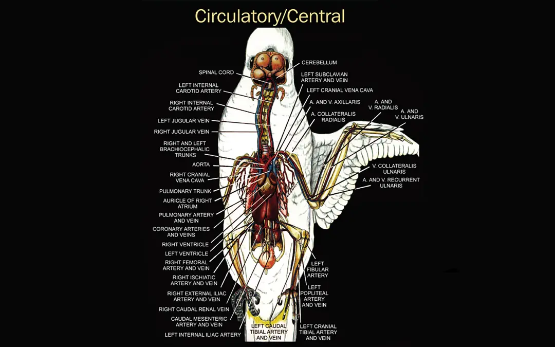
Psittacines follow a different anatomical pattern. Lesions can be present in any vessel but appear to subjectively concentrate in the aorta and brachiocephalic trunks rather than the coronary arteries. Myocardial infarction as a discrete event is uncommon.
Rather, the heart is affected indirectly, through the chronic pressure overload imposed by large-vessel stenosis, which drives ventricular hypertrophy, dilatation, and eventually systolic dysfunction. Pulmonary atherosclerosis can produce a separate pathway to right-sided congestive heart failure through pulmonary hypertension, presenting as coelomic effusion and hepatic venous congestion. Both pathways are possible in the same patient and population.
Nutrition
Nutrition is preventative, not curative. A complete, formulated diet reduces the dietary risk factors that contribute to atherogenesis, and transitioning away from seed-based feeding is the most impactful long-term intervention available for at-risk birds. Seed-based diets drive obesity and micronutrient deficiencies that promote vascular disease over time.
It is worth acknowledging that many companion parrots alive today were kept on seed-based diets for years or decades before adequate formulated options were widely available or before nutritional counseling became standard practice. These birds may have been accumulating vascular disease throughout that time. Improving the diet now is still worthwhile for long-term health, but it will not undo what has already developed. Atherosclerosis is not reversible, and neither owners nor clinicians should approach dietary change as a treatment for established disease.
- Harrison’s Adult Lifetime Fine or Coarse: Appropriate for most affected birds.
- Harrison’s High Potency: For those that are lean or nutritionally compromised, and for those who are transitioning from seed-based diets to a nugget for the first time.
For birds with unmanaged cardiovascular disease, dietary transition should not be initiated until the patient is clinically stable and under appropriate medical management. A bird in active cardiac decompensation is not a candidate for diet change. The metabolic and behavioral stress of transitioning a sick bird carries real risk and offers no acute benefit. This point is particularly important for owners to understand: changing the diet is not a treatment for a bird that is already ill, and it should never substitute for veterinary care.
How Avian Disease Differs from Dogs and Humans
Histopathologically, psittacine atherosclerosis closely resembles human disease, with shared features including foam cell formation and fibrous cap development. Clinically, however, the comparison breaks down quickly.
In humans, atherosclerosis drives coronary artery disease, and plaque rupture with acute coronary thrombosis is a defining clinical event. Validated staging systems and lipid-lowering treatment thresholds are built around this mechanism.
None of this maps directly onto birds:
- There is no equivalent staging system for avian cardiovascular disease.
- Coronary disease is not the dominant injury pathway.
- Acute events in birds typically reflect great vessel ischemia rather than coronary thrombosis.
- Statins, central to human cardiovascular management, depend on hepatic LDL receptor upregulation. That pathway does not translate cleanly to avian lipoprotein metabolism, and avian HDL-mediated reverse cholesterol transport remains poorly characterized. Research into whether statin use confers meaningful benefit in psittacines is ongoing, and to date extrapolation from human medicine has not been shown to produce a clear clinical benefit in birds.
Treatment and Expectations
Nothing reverses established atherosclerosis. Management goals are to slow progression, reduce cardiovascular burden, and preserve quality of life, but the appropriate strategy depends significantly on where the patient sits on the disease spectrum at the time of evaluation.
Birds with vascular disease but preserved cardiac function represent a different clinical situation than those with echocardiographic evidence of myocardial compromise, and both differ meaningfully from birds presenting in active congestive heart failure. Management should be tailored accordingly:
- Vascular changes with normal echocardiogram: Warrants consideration of vasodilators, monitoring, dietary optimization, and risk factor reduction, but may not yet require cardiac medications.
- Myocardial dysfunction identified: Medical support becomes appropriate. It is common in dogs to reach for pimobendan, but in parrots, the pathophysiology is different. Therefore, pimobendan use should be supported by poor contractility via echocardiogram prior to use.
- Active CHF with fluid accumulation: Furosemide for diuresis and coelomocentesis for respiratory relief may be necessary, though restraint of a decompensated bird carries real procedural risk and requires careful judgment.
Lesion location also matters: pulmonary atherosclerosis with right-sided CHF is managed differently than systemic vascular disease with left-sided dysfunction, and echocardiography is essential for distinguishing the two. For birds presenting in CHF regardless of the underlying distribution, prognosis is variable. Although some birds succumb early to their disease, many birds live happy lives many years past diagnosis as long as ongoing management is maintained.
Supplements
Omega-3 fatty acid supplementation, typically via fish oil, has a plausible mechanism of benefit through anti-inflammatory effects on the vascular endothelium. Available data in birds is limited and largely older, and a validated therapeutic dose for companion psittacines has not been established, but supplementation in at-risk birds on formulated diets is generally considered low-risk and reasonable. Several other supplements commonly used or investigated in human cardiovascular disease, including L-carnitine, CoQ10, and others, have not been meaningfully evaluated in parrots. Their efficacy and safety in this species are unknown, and they cannot be recommended on current evidence.
Environment and Activity
Encouraging physical activity is part of prevention, not treatment. For healthy at-risk birds, regular opportunities for flight, climbing, and foraging enrichment support cardiovascular fitness, weight management, and overall wellbeing. The principle is the same as in any species: start slow, build gradually, and pay attention to how the individual responds.
A bird that has been sedentary for years should not be pushed into sudden vigorous activity. If a bird appears to be struggling or is not tolerating increased activity well, scale back and reassess and present for veterinary evaluation. Exercise intolerance can be an early sign of cardiovascular disease.
In older birds, this can be complicated by comorbidities such as arthritis, which may limit mobility independently of cardiac status. Activity recommendations, enrichment choices, and the physical cage environment should all be tailored to the bird’s current level of mobility. Perch height, spacing, and cage layout should be adjusted as needed to reduce the risk of falls or trauma in birds whose balance or movement is compromised.
For birds that are decompensated or currently on cardiac medications, activity recommendations should come from the treating veterinarian. These patients require an individualized plan, and what is appropriate for one bird may not be safe for another, depending on the degree of cardiac involvement. Reducing chronic stressors, including social conflict, environmental instability, and excessive hormonal cycling, is a reasonable and low-risk component of long-term care for any bird with known cardiovascular risk.
References
- Girard C, Kummerfeld N, Pees M, Fehr M, Legler M. Characterization and classification of psittacine atherosclerotic lesions by histopathology, digital image analysis, transmission and scanning electron microscopy. Vet Sci. 2025;12(5):468. doi: 10.3390/vetsci12050468. PMID: 40431561.
- Wang WL, Chang FL, Yu PH. Chronic cor pulmonale secondary to pulmonary atherosclerosis in an African Grey parrot. BMC Vet Res. 2022;18(1):428. doi: 10.1186/s12917-022-03524-w. PMID: 36482409.
NOTE: Foundational epidemiologic data on psittacine atherosclerosis derives primarily from Beaufrere H et al., JAVMA 2013 (outside the 5-year window). Please confirm whether inclusion is acceptable before finalizing.
Disclaimer
This document reflects Harrison’s Bird Foods’ current understanding of atherosclerosis and cardiac disease in companion psittacines as of April 2026. Recommendations will be updated as the literature evolves. This is a summary of a complex topic; for comprehensive information, consult peer-reviewed literature and veterinary medical textbooks. This content does not constitute veterinary medical advice. Birds with medical concerns should be evaluated by a qualified avian veterinarian.